We use the most up-to-date technology to ensure the best eye care possible. Here are some of the different types of tests and equipment you may experience on a visit to our Practice.





Our Technologies
We are committed to bringing the best technology to our patients and being at the cutting edge, so we can render the best care.
We recently made 3 additions to our practice.
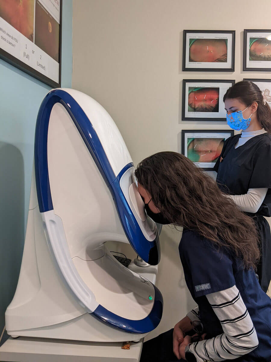
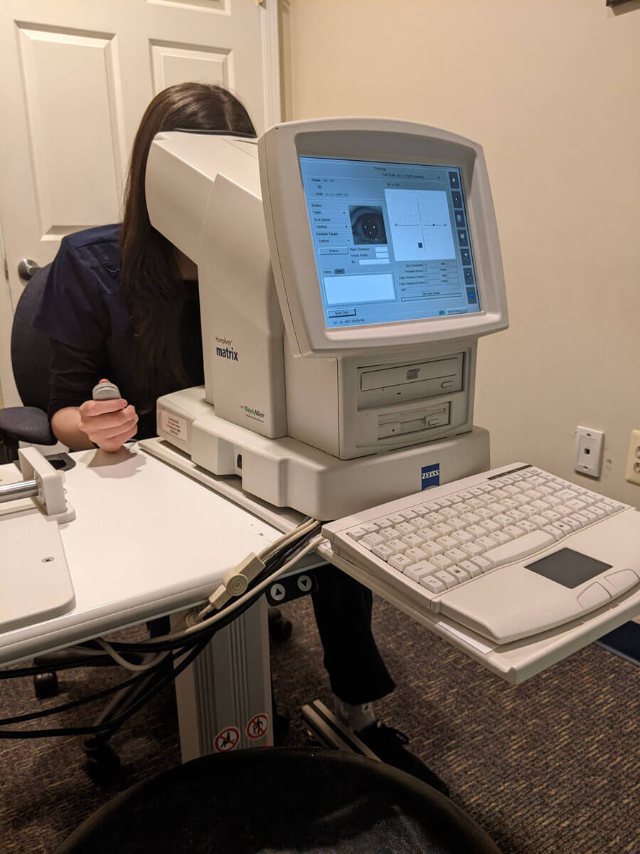
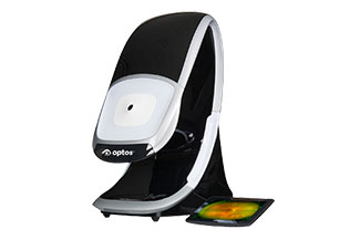
Virtual Field
The Virtual Field is a virtual reality headset instrument that measures your visual field; how you see to your sides, the periphery. It is very important in detecting and treating glaucoma, stroke, brain injury, and macular degeneration. It enables us to detect the early stages of disease and we can more easily monitor problems if they arise.
The benefits of this instrument is that it is faster, easier to use, and gives us even more valuable information than our prior automated visual field instrument. It's transportable to any room and it has audio that talks you through the test. It can even speak in over 30 languages.
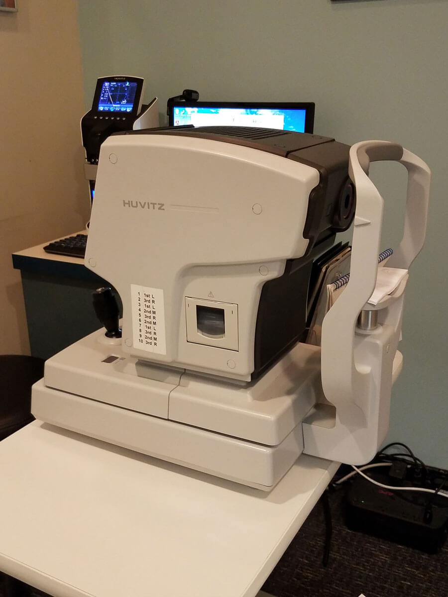
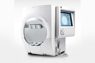
Pentacam
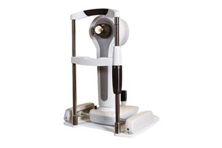
The Pentacam is a corneal tomographer. It takes virtual slices of the cornea, just as a CT or MRI takes virtual scans of the other parts of your body. It takes a precise and complete measurement and analysis of the cornea. This contact-free, hygienic measurement process takes only a few seconds. There is no radiation like that of a CT or MRI scan.
Within seconds the Pentacam gives us precise diagnostic information on the entire cornea, the front part of your eye. This instrument measures both the front and back surfaces of your cornea.
The whole cornea is just ½ a millimeter thick so being able to measure that front part of that ½ millimeter tissue and the back part of that thin tissue is truly amazing.
This information makes it possible to detect and monitor changes that are due to Keratoconus and Pellucid Marginal Degeneration.
If someone has had LASIK that has gone wrong, this instrument is able to very accurately measure the cornea so we can then design a special lens in order to restore sight.
This instrument captures thousands of data points of the cornea and we use this information when designing custom scleral lenses and when we design custom orthokeratology for you or your child’s eye, providing the best vision possible.
This is an amazing instrument and we are excited to bring you this technology.
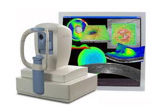
Optovue OCT
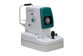
The iVue OCT (Ocular Coherence Tomography) takes 80,000 scans per second to assess the retina, optic nerve, and the cornea.
It takes virtual slices of the optic nerve and of the macula to help our patients who have glaucoma and those that have macular degeneration. It quantifies the thickness of the retina, nerve fiber layer, ganglion cell complex and the cornea and then compares that to thousands of people in the data bank.
The system tracks change and predicts trends. This instrument helps us diagnose and treat glaucoma, diabetic retinopathy, and so much more. We are so proud to bring this technology to you





Appointment times may vary so call us for availability.